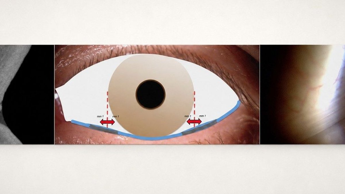
Fixed folds in the bulbar conjunctiva, termed LIPCOF, align parallel to the lower eyelid margin. These structures act as a clinical indicator associated with dry eye disease (DED) and modifications on the ocular surface.
Since LIPCOF grading is common in clinical settings and research, maintaining consistency is vital. However, differences in observation position can affect grading results, potentially reducing comparability across studies.
To address these issues, researchers investigated how observation position influences LIPCOF grading using two anterior segment imaging systems based on different OCT technologies.
Twenty young participants from the Cologne School of Optometry in Köln, Germany, took part in the study. For each participant, 21 vertical OCT line scans covering the meniscus area were captured in the LIPCOF region below both the nasal and temporal limbus using the Spectralis SD-OCT and Anterion SS-OCT devices.
For each device, the intended LIPCOF area directly below the limbus, as well as positions 1mm outward (towards the canthus) and 1mm inward (towards the pupil), were analyzed.
Read Also: Beauty products linked to rising eye health risks
A single masked examiner identified and graded visible LIPCOF from the OCT images in a randomized order.
Nasal LIPCOF grades from the Spectralis SD-OCT were significantly higher than those from the Anterion SS-OCT at the intended position and the 1mm outward position, differing by nearly half a grade in both cases (p<0.05). No significant differences were found at other locations (p>0.05).
With both devices, LIPCOF grades between intended and decentred positions correlated significantly in the temporal region but not in the nasal region. Decentration towards the canthus proved more forgiving than decentration towards the pupil.
Dr Stefan Bandlitz, head of the Cologne School of Optometry, led the research alongside students Franziska von der Höh and Andreas Ebner. Dr Bandlitz also serves as a visiting research fellow at Aston University, UK, specializing in contact lenses, anterior eye imaging, and the ocular surface.
